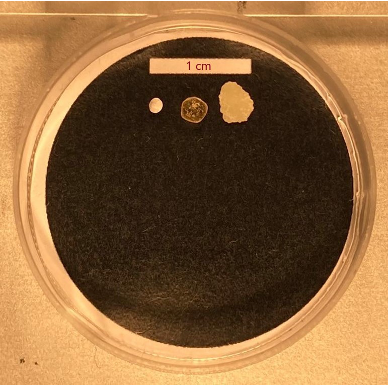
Every time we use the nets to sample the water we find a lot of interesting stuff, ranging from fish and critters to heaps of sargassum seaweed, and often a number of fragments which appear to be plastics. But are they really plastics though? Can we distinguish for example between a piece of plastic foil, and a translucent shell which might come from a shrimp or crab? Or taking it a step further, can we distinguish between different plastic polymers as well? As an example in Figure 1 you can see three particles in a petri dish. Can you spot the plastic particle(s)?
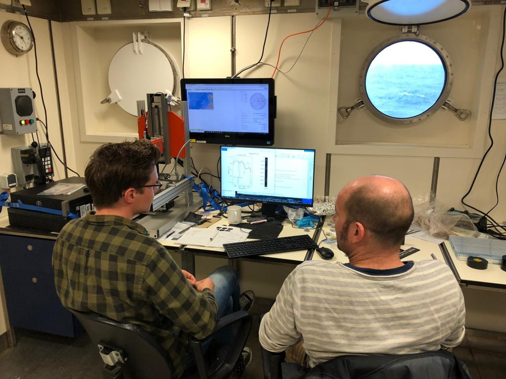
On board of the RV SONNE, we have a hyperspectral camera system (Fig. 2), measuring the near-infrared (NIR) spectrum reflected by the different particles. We first scan a long strip of these particles placed on a test-bed, which gives us some beautiful pictures like the one shown in Fig. 2. Image recognition software automatically detects the particles in the image. We then obtain the NIR spectrum for each particle. The spectra are compared to a library of reference materials. Different materials have for example different peaks in the spectrum.
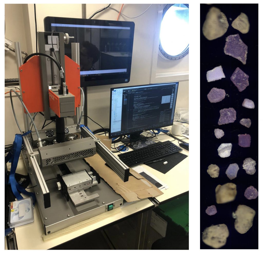
Right: Plastic particles placed on long strip. © M. Kaandorp
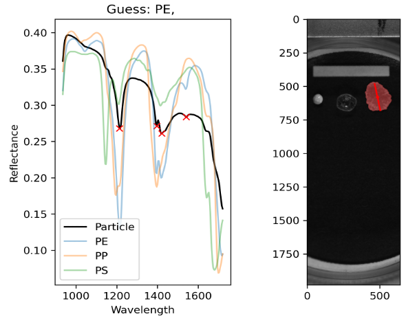
Figure 4 shows an example output from the software for the particle on the right in the petri dish. The left figure shows the NIR spectrum of the particle (black line), in the right figure the particle is marked in red by the image recognition software. After comparing the spectrum to the reference material library, it is classified to be likely a polyethylene (PE) particle. So far we see that most particles we obtained from the nets are polyethylene particles. This is not unexpected, since it is the most commonly found polymer in the environment. The other two particles in the petri dish are likely of biological origin: the particle on the left is a foraminifera, the particle in the middle is probably a piece of sargassum.